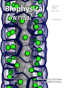
 Stem cells are fundamental building blocks for organ growth. They are cells that have not committed to doing a specific job and, therefore, can be directed by different signals to perform a wide range of tasks. The cover image shows the shapes of stem cells while they undergo the process of organ growth, in a computational model. The cells themselves are packed tightly, and so to reduce the amount of unused space in the organ they form a hexagonal arrangement. This isn’t unique to cells; if you pack oranges or balls on a shelf tightly you can see the same kind of packing. The type of packing reflects both the shape of the cells, and the amount of crowding in their environment.
Our simulations don’t generate these images automatically. When we want to analyse this type of system we take the raw data, typically in a text file, and use a specialized tool to view it as a 3D object. We did this using the tool VMD (http://www.ks.uiuc.edu/Research/vmd/) and rendering the cells as spheres. This is a very powerful way to show how the cells move and grow and is important for many types of analysis.
For this image we wanted to generate a different kind of visualization. We wanted an image that showed the cell packing unambiguously and more closely resembled the experimental microscopy data. To do this we performed a mathematical analysis—a Voronoi decomposition—to calculate the edges of the cells. For each cell we then rendered these faces as colored blue glass and drew in a small green sphere to show the center of the cell. This clearly shows how cells in the niche pack hexagonally, and the resulting image resembles the microscopy images much more closely. This makes direct comparison much simpler and can be used to validate the simulations.
Both the cover image and our article show what can be done using detailed computational models to understand organ growth. Although the work is focused on one specific system—the germline from the nematode C. elegans—both the model and the approach may have significant impacts beyond this system. The organ structure, a stem cell niche, is found commonly in many different systems, and just as tumors may grow in the C. elegans germline, mutations may cause human stem cell niches to develop into cancers. Similarly, our group at the MRC Cancer Unit, University of Cambridge, is looking to use the same methods and tools to model different pre-cancer and cancer systems. This includes studying detailed models of individual components and large biochemical networks in cells. The Fisher group at Microsoft Research and the Department of Biochemistry, Cambridge University, is using the same computational methods to model the molecular mechanisms underlying cancer (e.g., leukaemia, glioblastoma) as well as blood development.
For information on our research, visit the group’s website http://www.mrc-cu.cam.ac.uk/hall.html and blog http://drhallba.wordpress.com. For more information on some other work we have recently done on the structure of IKK-gamma, also visit the MRC Cancer Unit’s news section http://www.mrc-cu.cam.ac.uk/news.html. For information on the Fisher group, view http://research.microsoft.com/en-us/people/jfisher/ and recent press release related to their work on blood development http://research.microsoft.com/en-us/news/features/leukemia-drugs-computer-model.aspx.
- Benjamin A. Hall, Nir Piterman, Alex Hajnal, Jasmin Fisher