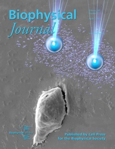
The cover image of Biophysical Journal (Volume 108, Issue 7) shows two chemoattractant-loaded microsource beads, which are being used to induce directed Jurkat cell migration. The image of the cell was obtained using SEM. The microsource beads, denoted by the two larger blue spheres, are fabricated using a solvent evaporation-spontaneous emulsion technique loaded with SDF-1α chemoattractant. The beads can be trapped and manipulated by optical tweezers, represented by the two beams of light, so that the chemoattractant molecules are released to form a concentration gradient that stimulates cell migration. The released molecules are denoted as a mass of small blue dots. Cell migration is involved in many fundamental biological phenomena, such as cancer, tissue repair, embryonic morphogenesis, and neural development. Cell migration is often driven by chemotaxis, where cells follow a gradient toward or away from a source of a specific chemical. For example, bacteria find food by swimming toward the highest concentration of food molecules, or inversely flee from poisons.
The technique represented by this cover image provides a powerful new tool to probe the mechanisms of chemotactic cell migration. In the associated study, this tool was used to reveal a novel cell migration model, which illustrates the relationship among the concentration gradient, the protrusion force, and the cell velocity. The theoretical prediction and the experimental results show that the motility of the cell increases when the gradient initially changes, indicating that the cell is sensitive to the initial external stimulation. However, both the cell velocity and protrusion force gradually stabilize when the gradient was higher than a certain value. This finding is novel and important for us to better understand cell migration.
There are still many outstanding questions about chemotaxis and cell migration. The approach shown here provides an effective tool to study cell migration in a controllable, observable, and flexible way. The data available from this approach are also relevant to the development of migration-based biomedical applications, such as targeted therapy, stem cell therapy, and tissue engineering. Further information on our research can be found at
: http://www.cityu.edu.hk/mbe/medsun/
– Hao Yang, Xue Gou, Yong Wang, Tarek M Fahmy, Anskar Y-H Leung, Jian Lu and Dong Sun